Introduction: A Turning Point in AI Clinical Research
In the rapidly advancing world of healthcare, artificial intelligence (AI) has moved from being a futuristic concept to a practical tool reshaping medical practice and clinical research. One of the biggest bottlenecks in biomedical studies is the manual annotation of medical images — a task that can take weeks or months for researchers and radiologists.
Recently, researchers at the Massachusetts Institute of Technology (MIT) unveiled a groundbreaking AI system that automates this critical but time-consuming process. The tool not only accelerates clinical research but also enhances accuracy, reproducibility, and scalability in analyzing biomedical data.
This innovation is more than a technical upgrade; it represents a paradigm shift in how medical science approaches research efficiency, with implications for disease discovery, treatment evaluation, and the future of personalized medicine.
The Challenge of Manual Image Annotation
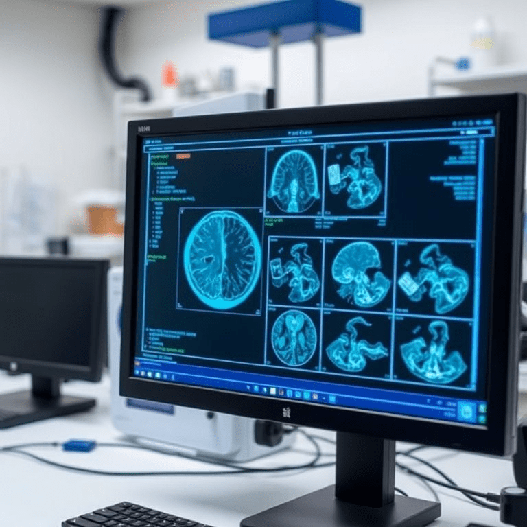
Medical image annotation is a cornerstone of clinical research. Researchers frequently analyze MRI scans, CT scans, ultrasound images, or microscopic biomedical data to study diseases ranging from Alzheimer’s to cancer.
However, annotating these images — a process known as segmentation — is labor-intensive:
- Time-consuming: A single dataset with thousands of images may require hundreds of hours of expert attention.
- Prone to human variability: Different researchers may annotate the same image differently, creating inconsistencies.
- Resource-intensive: Requires highly trained professionals such as radiologists, pathologists, or biomedical engineers.
For example, mapping changes in the hippocampus to study Alzheimer’s disease progression involves outlining each hippocampus across hundreds of brain scans. This task, while critical, drains valuable time that could otherwise be spent on analysis, hypothesis testing, or clinical trials.
How MIT’s AI Tool Works
The MIT team developed an AI tool that uses interactive machine learning to streamline annotation. Instead of manually outlining every detail, researchers can click, scribble, or draw boxes on biomedical images.
Key Features:
- Progressive Learning
- With each annotation, the AI learns and requires fewer human inputs.
- Over time, it can segment new images independently.
- Dataset-Level Efficiency
- Unlike traditional AI models that require manual labeling of each image, this system learns across the dataset, making predictions for all subsequent images.
- Adaptability
- Works across various imaging modalities (MRI, CT, histopathology).
- Can adapt to specific research datasets without retraining from scratch.
- Reduced Errors
- Continuous feedback loop with researchers minimizes false annotations.
According to MIT’s developers, this reduces annotation time by up to 80%, enabling teams to complete studies weeks faster than before.
Why This Matters for Clinical Research
1. Faster Medical Discoveries
Clinical research often faces delays due to data preparation bottlenecks. By accelerating annotation, this tool ensures faster turnaround times for publishing results and testing treatments.
2. Improved Accuracy
AI’s consistency minimizes inter-observer variability, creating more reproducible and standardized datasets. This reliability is crucial for multi-center studies where data consistency can determine trial success.
3. Cost Reduction
Medical research is expensive, with annotation often accounting for 30–40% of project costs. Automating this step frees up funds for other phases of research, including drug trials or patient recruitment.
4. Scalability
As the biomedical field moves into big data territory with genome sequencing, 3D imaging, and personalized diagnostics, scalability becomes critical. MIT’s tool ensures researchers can handle large datasets without scaling costs proportionally.
Applications in Real-World Healthcare
This AI annotation tool has vast potential applications:
🧠 Neurology Research
- Mapping brain structures for Alzheimer’s, Parkinson’s, and epilepsy studies.
- Accelerating large-scale brain mapping projects like the Human Connectome Project.
🩺 Oncology
- Rapid tumor detection and measurement in CT/MRI scans.
- Annotation of microscopic images for cancer cell segmentation.
🧬 Genetics & Pathology
- Assisting pathologists in analyzing genomic imaging data.
- Reducing the time to identify cell mutations in tissue samples.
🫁 Pulmonology & Cardiology
- Measuring lung damage in COPD patients.
- Automating detection of arterial plaque for cardiovascular disease risk.
🦠 Infectious Disease Research
- Mapping tissue damage in diseases like COVID-19 or tuberculosis.
- Speeding up vaccine development research by providing annotated biological data.
Ethical Considerations and Limitations
While promising, the integration of AI in clinical research also raises challenges:
- Bias in Training Data: If initial datasets are biased, AI could make skewed predictions.
- Data Privacy: Biomedical images often contain sensitive patient data, requiring strict adherence to HIPAA and GDPR standards.
- Overreliance on AI: Researchers may risk overtrusting AI predictions without adequate validation.
- Regulatory Approval: Widespread use in clinical trials requires FDA or EMA approvals, which may slow adoption.
Expert Opinions
Dr. James Lee, a radiology researcher at Harvard Medical School, commented:
“Annotation has always been one of the most painful parts of biomedical research. MIT’s tool doesn’t just save time — it ensures consistency across large datasets. That’s a game changer for clinical trials.”
Similarly, Dr. Ananya Desai, a biomedical engineer at AIHealthTech, said:
“The scalability of this system is impressive. For the first time, we’re looking at AI that can handle the data tsunami in medical research without compromising accuracy.”
The Future of AI in Clinical Research
The MIT tool is part of a broader trend where AI and machine learning are becoming indispensable in medicine. Looking forward, we can expect:
- Integration with Electronic Health Records (EHR): Linking annotated imaging data with patient histories for personalized treatment.
- Real-Time Clinical Applications: Doctors could use similar systems during surgeries or diagnostics for instant segmentation.
- AI-Driven Clinical Trials: Faster patient selection, monitoring, and analysis.
- Collaborative AI Networks: Institutions sharing annotated datasets securely to accelerate global medical discoveries.
Conclusion
MIT’s new AI tool is not just a technical advancement — it’s a transformational step in medical science. By automating biomedical image annotation, it tackles one of the biggest hurdles in clinical research: time-consuming manual work.
With the potential to accelerate disease research, cut costs, and improve accuracy, the tool positions AI as a central driver in the next era of healthcare innovation. While challenges around ethics, regulation, and data security remain, the trajectory is clear: AI will be at the heart of clinical research in the future.